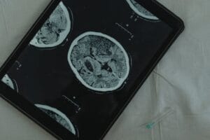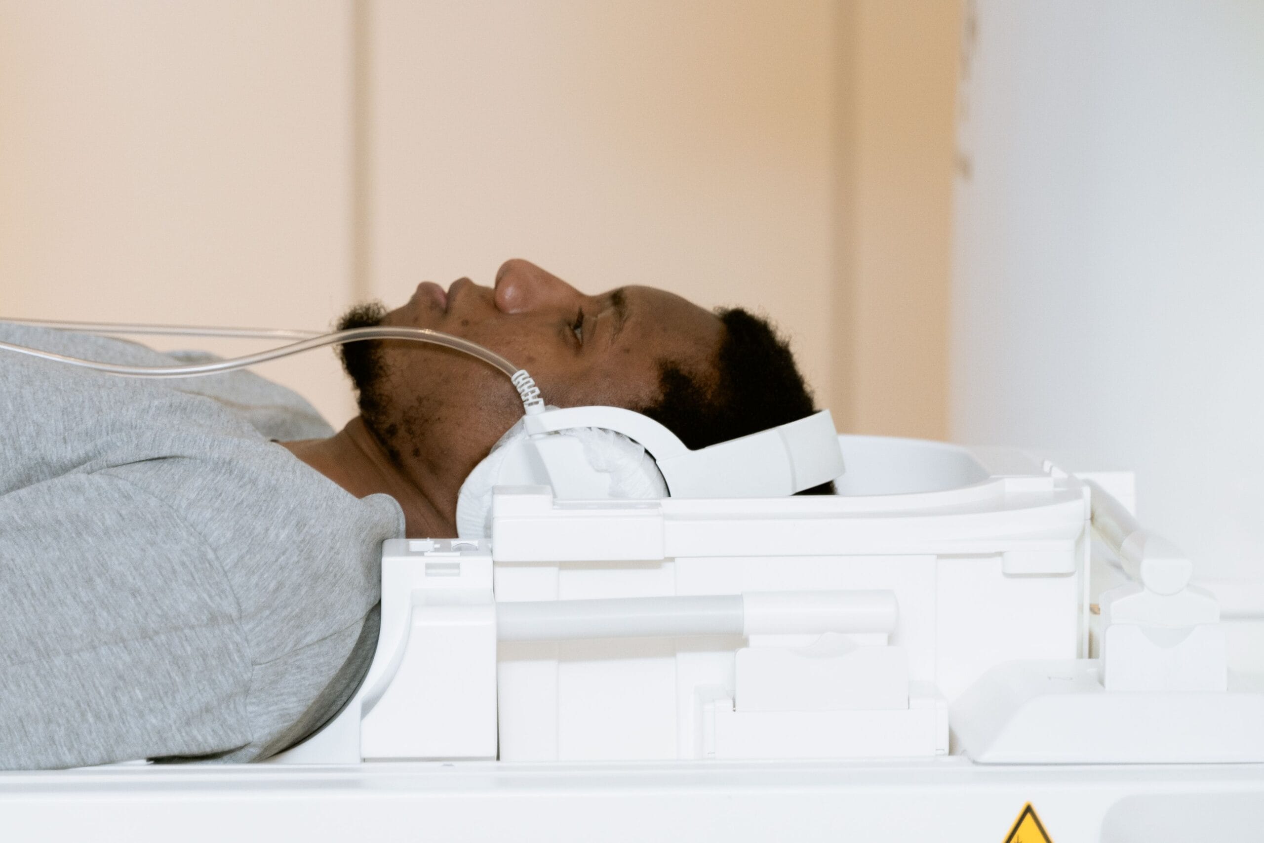
You’ve been told you need a scan, or you’re weighing up one your GP mentioned against another you’ve read about. It’s the question every radiologist hears, and the honest answer is not the one most articles will give you. Neither scan wins outright. A CT and an MRI see the body in completely different ways, which is why they exist alongside each other, not in place of each other.
The choice comes down to what needs to be seen. This article explains how each scan works, what each one shows that the other cannot, why field strength matters when it comes to MRI, and how doctors decide between them for problems ranging from a suspected fracture to an unexplained headache.
CT scan vs MRI: the difference at a glance
CT scan
Sees with: X-rays
Scan time: minutes
Radiation: low, controlled dose
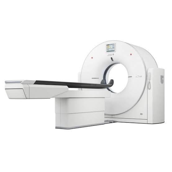
Feels like: an open ring, quick and quiet
Strongest at: bone, lungs, bleeding, speed
MRI scan
Sees with: magnet and radio waves
Scan time: 20 to 60 minutes
Radiation: none
Feels like: an enclosed tunnel, noisy, longer
Strongest at: brain, spine, joints, soft tissue
What is a CT scan?
A CT scanner is essentially an X-ray machine that circles you. As the beam rotates, it captures hundreds of images from different angles, and a computer stacks them into cross-sectional slices of your body. From the table, the whole thing feels unremarkable. You lie inside an open ring, hold your breath once or twice, and the scan itself is over in minutes.
That speed is exactly why CT is the workhorse of emergency medicine. It shows bone, the lungs, internal bleeding and problems in the abdomen quickly and reliably. If you arrive at A&E after an accident, a CT scan is usually the first detailed test you’ll have. The NHS overview of CT scans walks through the procedure step by step.
What is an MRI scan?
An MRI takes a completely different route. Instead of X-rays, it surrounds you with a strong magnetic field, sends radio waves through your body, and listens to the signals coming back. Different tissues respond differently, and from those responses the machine builds its images. No ionising radiation is involved at any stage.
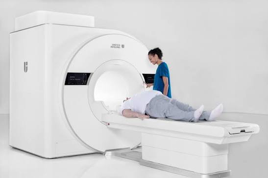
The trade-off is time and comfort. A typical MRI takes anywhere from 20 minutes to an hour inside a tunnel-shaped scanner that knocks and hums loudly while you hold still. In return, it produces soft tissue detail that nothing else can match, which is why it’s the preferred scan for the brain, spinal cord, ligaments, joints, and organs such as the prostate and liver. The NHS guide to MRI scans covers what to expect on the day.
Not all MRIs are the same: 1.5T and 3T ultra wide-bore
Most articles stop at “MRI” as if there’s only one kind. There isn’t. The two most common MRI scanners in clinical use are 1.5 Tesla and 3 Tesla, and the Tesla figure refers to the strength of the magnetic field. A 3T scanner has twice the field strength of a 1.5T, which produces sharper images and can shorten the time you spend in the scanner without losing detail. For anyone claustrophobic, the shorter time alone is a meaningful upgrade.
The other variable is the size of the opening you lie inside, called the bore. Standard scanners have a bore around 60cm across. Ultra wide-bore designs open the tunnel to 70cm, and the widest available now reach 75cm. That extra width doesn’t sound like much on paper. But it changes the experience significantly for anyone who dislikes enclosed spaces, and it also expands who can be scanned. Top-of-class ultra wide-bore machines can accommodate patients up to around 310 kg, opening MRI to people who previously couldn’t fit inside a standard scanner at all. Claustrophobia is a well-documented reason people delay, decline, or cut short an MRI, and ultra wide-bore scanners were designed specifically to address that.
3T ultra wide-bore, in plain terms
Sharper images: the magnetic field is twice as strong as a 1.5T scanner, so more fine detail comes through.
Shorter scan: less time inside the machine for the same information.
Wider opening: the bore reaches 75cm across at the top of the class, nearly a third wider than a standard 60cm scanner.
Higher weight capacity: can accommodate patients up to around 310 kg, well beyond the limit of a standard MRI.
Making MRI more bearable: the comfort features
The top of this class combines field strength, bore width, and a set of comfort features designed around the parts of an MRI people find most difficult. AI-guided imaging shortens the scan by identifying the best sequences automatically. Quiet-scan technology reduces the acoustic noise by close to half, from the loud knocking most people remember to something much closer to a hum. And above your head, a backlit ceiling image, often a starlit sky, gives the eye something calm to rest on for the length of the scan.
Why fewer people now need sedation for MRI
Together, these features mean many people who used to need sedation to complete an MRI can now do so awake, calmly, and often with a companion nearby. That includes children. Purpose-built infant and paediatric coils allow scans to be performed without sedation, while a parent stays in the room throughout. The same combination of speed and image quality that helps with planned imaging also has a role in accident and emergency care. Fast, clear pictures can change the treatment path.
An ultra wide-bore 3T MRI at this level combines every advantage in one machine. Sharper detail, shorter scan time, a wider opening, and a design deliberately built around the person going into it. It represents the most advanced form of routine clinical MRI currently in use, and it’s the class of scanner we cover in detail in our guide to 3T full body MRI. If field strength or comfort matters to you, this is the class of scanner worth asking about before you book.
What does an MRI show that a CT scan cannot?
MRI’s real gift is telling apart soft tissues that look almost identical on a CT image. A tendon from the fluid around it. Healthy liver from something that shouldn’t be there. In the first hours after a stroke, an MRI can reveal injured brain tissue while a CT scan may still look completely normal. That kind of early detail matters, because it changes how the stroke is treated.
MRI is also the scan of choice for spinal cord compression, torn ligaments and cartilage damage, and subtle changes deep in the brain. It does more of the explaining, too. A CT scan may show that something is there. An MRI can often say what that something is likely to be. This is why doctors sometimes order an MRI after a CT scan, not instead of one.
What does a CT scan show better than an MRI?
Bone, first and foremost. CT shows fractures, bone density changes and skull injuries with a sharpness MRI simply cannot match. It’s also the stronger scan for the lungs, where it picks up small nodules and early signs of disease that other imaging can miss. And for fresh internal bleeding, CT finds it quickly, which in an emergency is everything.
Speed settles many decisions too. A CT takes minutes rather than the best part of an hour, so it wins whenever time is critical, or when someone cannot lie still for long. And for people whose implants or medical devices make an MRI unsafe, CT is often the practical answer.
CT scan vs MRI: which one for which problem?
Your doctor makes the final call, because your history and symptoms shape the decision. But the patterns below hold in most cases, and knowing them helps the conversation.
A suspected broken bone
An emergency after an accident
A detailed look at the lungs
Unexplained headaches or neurological symptoms
Back pain with nerve symptoms such as sciatica
A joint, ligament or cartilage injury
A closer look at the prostate, liver or breast
CT scan vs MRI: so which is better?
Neither, because the question is the wrong way round. The two scans are colleagues, not rivals. Radiologists don’t start with the machine. They start with the clinical question: what needs to be seen, how urgently, and in which part of the body? Once those three things are clear, the scan chooses itself.
A suspected lung problem points to CT. An unexplained neurological symptom points to MRI. And a thorough look at the whole body often needs both, because each covers the other’s blind spots.
Clinical perspective
Dr Vidhi Panchal
Radiologist
The question in imaging is never which scanner is more advanced. It’s which combination of images gives the clearest answer to the question your doctor is actually trying to answer. That judgement, not the technology on its own, is what turns a scan into a decision you can act on.
Where does a PET CT scan fit in?
A PET CT pairs a CT scanner with a small amount of radioactive tracer that highlights how tissue is behaving. CT and MRI show how the body looks. PET CT shows what it’s doing. It sits alongside the other two, not in place of them, which is why it appears most often in cancer assessment, where knowing whether a spot is active can tell the whole story. Imaging is only one part of a complete check, and our overview of what a full body MOT includes covers the wider picture.
CT scan vs MRI: what nobody explains about radiation, dye, and the tube
Radiation is the most common worry, so it deserves a straight answer. A CT scan uses a low, carefully controlled dose of X-rays. According to the NHS, the exposure from a single scan is broadly comparable to the natural background radiation you’d absorb from the environment over months to a few years, depending on the type of scan. Doctors only recommend one when the benefit clearly outweighs that small risk. MRI, for its part, uses no ionising radiation at all.
Both scans sometimes use a contrast dye to make certain structures stand out. CT uses an iodine-based dye. MRI uses a gadolinium-based one. The body clears both naturally, usually within a day or two, and clinics often check kidney function beforehand as a precaution.
What the tube actually feels like
Comfort divides the two more sharply. A CT scanner is an open ring, and the scan is over quickly, so very few people find it difficult. An MRI is longer, louder, and more enclosed. If you’re the sort of person who dislikes small spaces, the tunnel is what tends to worry people most. This is where ultra wide-bore 3T MRI comes into its own, with a much larger opening and a shorter scan time. Tell the radiographer beforehand. You can also ask about eye masks, headphones, or going in feet first, all of which help more than people expect.
CT scan vs MRI: frequently asked questions
Is a CT scan the same as an MRI? +
No. Both produce detailed images of the inside of the body, but they work in completely different ways. A CT scan uses X-rays to build cross-sectional slices. An MRI uses magnetic fields and radio waves. That difference is why each suits different parts of the body and different clinical questions.
What’s the difference between a 1.5T and a 3T MRI? +
The number refers to the strength of the magnetic field. A 3 Tesla scanner has twice the field strength of a 1.5 Tesla, which produces sharper images and generally shortens the time you spend in the machine. Not every clinical question needs 3T, but for detailed imaging of the brain, spine and small joints, higher field strength usually helps.
Do CT scans give off a lot of radiation? +
A CT scan uses more radiation than a standard X-ray, but the dose is low and carefully controlled. According to the NHS, the exposure from a single scan is broadly comparable to the natural background radiation you’d absorb over months to a few years, depending on the scan. Doctors only recommend a CT scan when the benefit clearly outweighs that small risk.
Is contrast dye harmful to the body? +
For most people, no. CT scans sometimes use an iodine-based dye, and MRI scans a gadolinium-based one. The body clears both naturally, usually within a day or two. Serious reactions are uncommon. Clinics still tend to check kidney function beforehand, because the kidneys do most of the work in removing contrast from the body.
Getting a scan: the practical questions
Why won’t my doctor order me an MRI? +
In most cases it’s because an MRI wouldn’t change your diagnosis or treatment. GPs follow clinical guidelines that reserve MRI for situations where the result will genuinely alter the next step. NHS scanning capacity is also limited, so referrals are prioritised by clinical need. If you feel your concern has not been fully explored, ask your GP to explain the reasoning, or ask about private options.
Can I ask my GP to refer me for an MRI scan? +
Yes, you can always ask, though the decision rests with your GP and depends on clinical justification. NHS MRI referrals go through a needs-based prioritisation system. Private MRI scans are widely available on a self-pay basis if you want to move sooner.
When an MRI is difficult or unsafe
What triggers panic during an MRI, and what actually helps? +
The enclosed tunnel, the noise, and the need to lie still for 20 to 60 minutes are the usual triggers. What helps: telling the radiographer beforehand so they can talk you through it, using the earplugs or headphones provided, closing your eyes for the whole scan, and asking whether you can go in feet first. Ultra wide-bore scanners have a much larger opening and shorter scan times, which many people find far easier.
Can children have an MRI without sedation? +
Sometimes, yes. Sedation is often used when a child can’t stay still long enough for a clear image. Newer wide-bore scanners with dedicated paediatric coils and shorter scan times have made it possible to scan more children awake, often with a parent staying in the room during the scan. Whether it’s offered depends on the scanner, the age of the child, and the type of imaging needed, so ask the hospital or clinic beforehand.
Who should not have an MRI scan? +
Anyone with certain metal implants or medical devices needs a safety assessment first. Older pacemakers, some surgical clips and implants, and metal fragments in the body can make MRI unsafe, so always tell staff about any device or implant before the scan. In these cases, a CT scan is often the safer option, which is one more reason both technologies remain essential.
Understanding what each scan does is one thing. Bringing them together into a full picture of your health is another.